

DOI: 10.5281/zenodo.23674455 · UDC: [616.366-003.7-002.1+616.36-008.5]-089
Background. Obstructive jaundice associated with hepatopancreatobiliary disease is a clinically important condition that may accompany gallstone-related inflammatory complications. Management is challenging because gallstone disease, infection or inflammation, and biliary obstruction may need to be addressed simultaneously.
The purpose of the study. To evaluate the outcomes of the sequential approach and describe the management strategy used in patients with gallstone disease complicated by infection and cholestatic jaundice.
Methods. This retrospective descriptive study presents the sequential management of 67 adult patients with acute calculous cholecystitis and/or cholangitis associated with cholestatic jaundice, treated at the “St. Archangel Michael” Municipal Clinical Hospital, Chișinău, Republic of Moldova, from 2006 to 2017. The dataset was derived from a doctoral research project; some findings had previously been reported only partially. Treatment strategies were individualized according to the clinical presentation and severity of biliary infection. The research received approval from the hospital Ethics Committee in 2006.
Results. The age range of participants was 23 to 82 years with an average age of 63.46 ± 14.22 years, and included 44 (65.7%) women and 23 (34.3%) men. In 16 patients with a simple form of acute cholecystitis and jaundice, caused by choledocholithiasis (3 cases) and stenosis of the sphincter of Oddi (13 cases), the endoscopy-laparoscopy technique was applied together with papillosphincterotomy (PST) within the first 48–72 hours followed by laparoscopic cholecystectomy within 3–4 days after biliary drainage. In acute destructive cholecystitis associated with jaundice, caused by choledocholithiasis – 13 cases, stenosis of the sphincter of Oddi – 6 cases and severe cholangitis – 12 cases, the techniques applied were: 1) Laparoscopy – endoscopy with laparoscopic cholecystectomy with Halsted drainage – 10 patients, with subsequent PST and stone removal when indicated, including during readmission after fistulography, 2) Laparotomy – endoscopy: classical cholecystectomy, choledochotomy with stone removal and Kehr drainage – 21 patients, fistulography on the 5th–7th day postoperatively followed by PST with stone removal in 17 patients. In 7 cases of residual choledocholithiasis associated with severe cholangitis and jaundice, PST with stone extraction was performed, in 3 cases associated with endoscopic nasobiliary drainage, and in 1 case – with placement of an internal biliary stent. In 13 cases of Mirizzi Syndrome, endoscopy-laparotomy techniques were applied, in the 1st stage – endoscopic retrograde cholangiopancreatography (ERCP) with PST – 10 patients, in 3 patients – endoscopic nasobiliary drainage. In the second stage, biliary tract reconstruction interventions were performed depending on the topography of the fistula, assessed during intraoperative cholangiography. Eight complications were reported in the 67-patient cohort, with no deaths.
Conclusions. In the studied cohort, the timing and type of intervention were individualized according to the morphological form of the disease and the patient's clinical condition, with biliary decompression prioritized in the first stage of sequential management.
Introducere. Icterul obstructiv asociat cu patologia hepatopancreatobiliară este o afecțiune clinică importantă care poate însoți complicațiile inflamatorii ale litiazei biliare. Managementul reprezintă o provocare deoarece litiaza biliară, infecția sau inflamația și obstrucția biliară ar putea necesita o abordare simultană.
Scopul studiului. Evaluarea rezultatelor abordării secvențiale și descrierea strategiei de management utilizate la pacienții cu litiază biliară complicată cu infecție și icter colestatic.
Metode. Acest studiu descriptiv retrospectiv prezintă managementul secvențial al 67 de pacienți adulți cu colecistită acută litiazică și/sau colangită asociată cu icter colestatic, tratați la Spitalul Clinic Municipal „Sfântul Arhanghel Mihail”, Chișinău, Republica Moldova, în perioada 2006-2017. Setul de date a fost derivat dintr-un proiect de cercetare doctorală; unele constatări au fost raportate anterior doar parțial. Strategiile de tratament au fost individualizate în funcție de tabloul clinic și de severitatea infecției biliare. Cercetarea a primit aprobarea Comitetului de Etică al spitalului în anul 2006.
Rezultate. Vârsta participanților a fost cuprinsă între 23 și 82 de ani, cu o medie de 63,46 ± 14,22 ani, lotul incluzând 44 (65,7%) femei și 23 (34,3%) bărbați. La 16 pacienți cu formă simplă de colecistită acută și icter, cauzată de coledocolitiază (3 cazuri) și stenoza sfincterului Oddi (13 cazuri), s-a aplicat tehnica endoscopie-laparoscopie împreună cu papilosfincterotomia (PST) în primele 48–72 de ore, urmată de colecistectomie laparoscopică la 3–4 zile după drenajul biliar. În colecistita acută distructivă asociată cu icter, cauzată de coledocolitiază – 13 cazuri, stenoza sfincterului Oddi – 6 cazuri și colangită severă – 12 cazuri, tehnicile aplicate au fost: 1) Laparoscopie – endoscopie cu colecistectomie laparoscopică cu drenaj Halsted – 10 pacienți, cu PST ulterioară și extragerea calculilor atunci când a fost indicat, inclusiv în timpul reinternării după fistulografie; 2) Laparotomie – endoscopie: colecistectomie clasică, coledocotomie cu extragerea calculilor și drenaj Kehr – 21 pacienți, fistulografie în ziua 5–7 postoperator urmată de PST cu extragerea calculilor la 17 pacienți. În 7 cazuri de coledocolitiază reziduală asociată cu colangită severă și icter s-a efectuat PST cu extragerea calculilor, în 3 cazuri asociată cu drenaj nazobiliar endoscopic, iar într-un caz – cu montarea unui stent biliar intern. În 13 cazuri de Sindrom Mirizzi s-au aplicat tehnici de endoscopie-laparotomie: în etapa I – colangiopancreatografie retrogradă endoscopică (ERCP) cu PST – 10 pacienți, la 3 pacienți – drenaj nazobiliar endoscopic. În etapa a II-a s-au efectuat intervenții de reconstrucție a căilor biliare în funcție de topografia fistulei, evaluată în timpul colangiografiei intraoperatorii. S-au raportat 8 complicații în cohorta de 67 de pacienți, fără niciun deces.
Concluzii. În cohorta studiată, momentul și tipul intervenției au fost individualizate în funcție de forma morfologică a bolii și de starea clinică a pacientului, decompresia biliară fiind prioritizată în prima etapă a managementului secvențial.
The medical and surgical approach to a patient with jaundice remains a challenge for the practicing surgeon. Obstructive jaundice syndrome in diseases of the hepato-pancreatobiliary system has an incidence of 2.6%–23.7% [1], being characteristic of both benign and malignant pathologies. Statistics reveal a significant number of surgical interventions on the extrahepatic biliary tract are performed for gallstone disease and icterus, which is considered the most common cause of benign mechanical jaundice [1, 2]. Known as surgical jaundice, the icteric syndrome in complications of gallstone disease is primarily caused by secondary choledocholithiasis. However, jaundice also frequently occurs during inflammatory complications, such as acute cholecystitis, cholangitis, pancreatitis, and reactive hepatitis [1, 2, 3, 4]. Cholestatic jaundice is currently considered a complex, multifactorial pathogenic process that serves as the primary etiological basis for the superimposition of microbial factors, leading to serious functional disturbances with severe repercussions for the liver [2, 3]. Studies demonstrate that prolonged cholestatic jaundice is a major cause of hepatic homeostasis disruption, progressively inducing hepatocellular insufficiency associated with high morbidity and mortality. A patient with gallstone complications associated with jaundice presents a difficult medical emergency. The difficulty of managing such patients results from the need to address three problems simultaneously: gallstone disease, the associated inflammatory complication, and cholestatic jaundice. Furthermore, cholestatic jaundice may develop both in young patients with an uncomplicated clinical course and in elderly patients with severe comorbidities, creating a dual challenge in diagnosis and management [2, 4]. Consequently, the medical-surgical management requires prompt intervention using a well-defined, selective diagnosis and therapeutic strategy that is strictly tailored to the morphological form of the disease and the physiological condition of the patient [5, 6].
The primary objectives of surgical intervention in these patients are biliary decompression, improvement of bile flow, and the resolution of both biliary lithiasis and inflammatory complications [5, 6]. Given the severity and unpredictable course of the disease, numerous publications emphasize that the ideal intervention should be technically straightforward for the specialist and well-tolerated by the patient, while being associated with a low rate of postoperative complications. Furthermore, it should employ minimally invasive techniques that are accessible to a wide range of medical institutions [6].
The current interventional options for managing gallstone disease associated with infectious complications and jaundice are diverse. They include the classical open single-stage approach, the single-stage laparoscopic approach, sequential endoscopy followed by laparoscopy, sequential laparoscopy followed by endoscopy; and the simultaneous laparo-endoscopic approach [5, 6, 7].
Controversies regarding the treatment of complicated cholelithiasis associated with mechanical jaundice are strongly influenced by socio-economic factors, as some cutting-edge techniques remain inaccessible to most patients or clinics [6, 7, 8, 9]. Despite numerous studies on the subject, there is still no universally accepted consensus regarding the optimal timing, type and extent of surgical intervention.
The purpose of the study. To evaluate the outcomes of the sequential approach and describe the management strategy used in patients with gallstone disease complicated by infection and cholestatic jaundice.
This paper presents a comprehensive and detailed description of the results of sequential treatment for inflammatory complications of gallstone disease associated with cholestatic jaundice - results previously published only partially and in a fragmented manner in certain articles. The material was selected from a doctoral thesis that outlined a differentiated therapeutic approach to cholestatic jaundice, tailored to the specific complication present in the context of gallstone disease. In accordance with the proposed purpose, this study included 67 patients presenting with acute calculous cholecystitis, cholangitis and cholestatic jaundice. Patients in this group underwent sequential surgical treatment, employing various strategies and techniques, at the "St. Archangel Michael" Municipal Clinical Hospital in Chișinău, Republic of Moldova, between 2006 and 2017. The research was conducted with approval from the Ethics Committee of the “St. Archangel Michael” Municipal Clinical Hospital in 2006. The data were collected prospectively, as part of the doctoral thesis, and analyzed retrospectively. The severity of the inflammatory process was retrospectively reclassified according to TG18.
Study inclusion criteria:
Adults aged >18 years; acute calculous cholecystitis and/or cholangitis associated with cholestatic jaundice; verbal consent to participate.
Study exclusion criteria:
Age <18 years; gallstone disease with hepatocellular jaundice; gallstone disease with jaundice and oncological pathology confirmed preoperatively, intraoperatively, or postoperatively; verbal refusal to participate.
Data were compiled and processed using Microsoft Excel (Microsoft Office 2010) and GraphPad software. The analysis was primarily descriptive and included means with standard deviations for continuous variables and absolute and relative frequencies for categorical variables. Because no inferential comparisons are reported in the Results, statistical significance testing is not used to support the conclusions of the present manuscript.
The age of the patients ranged from 23 to 82 years, with an average age of 63.46 ± 14.22 years. The study group included 44 (65.7%) women and 23 (34.3%) men. The demographic composition of the study group, based on inclusion questionnaires.
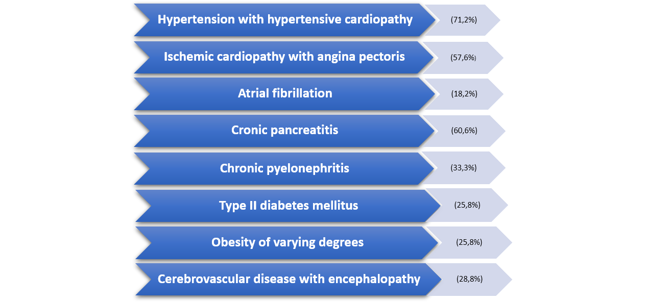
The comorbidities in the study groups were predominantly cardiovascular and digestive, 32% of patients presenting multiple comorbidities in one patient (Figure 1).
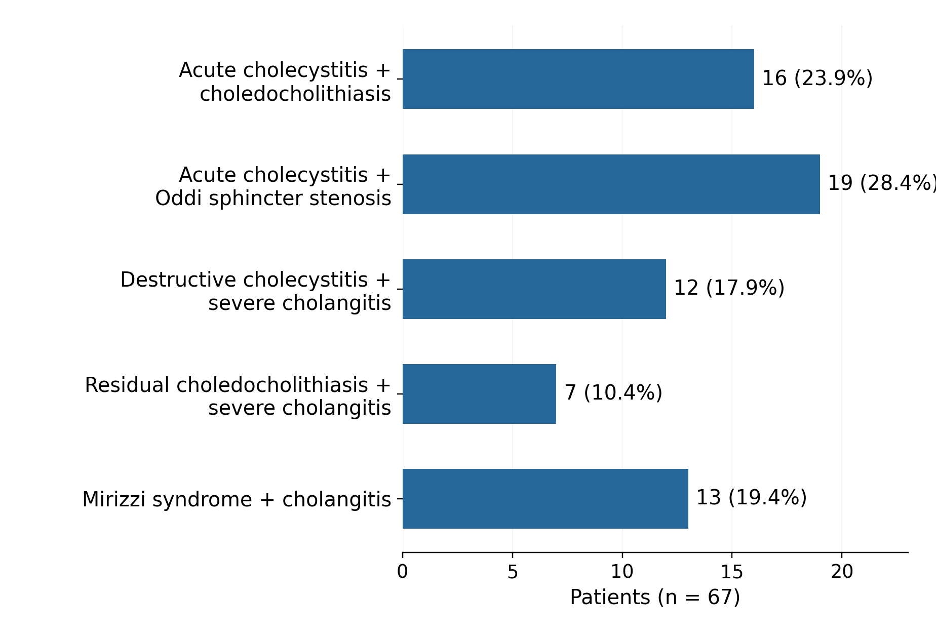
Patients were evaluated based on the anatomical localization of the cause of jaundice, alongside the diagnostic criteria for acute cholecystitis (A+B+C) and cholangitis (Gr.1; Gr.2; Gr.3) according to the Tokyo Guidelines 2018 (TG18) [11, 12]. Sphincter of Oddi stenosis was diagnosed visually via duodenoscopy and confirmed by MR cholangiography in 6 cases and ERCP in 13 cases. The study included several groups of patients with cholestatic jaundice caused by infectious complications of gallstones: 16 (23.9%) patients with acute calculous cholecystitis (AC) associated with choledocholithiasis (CL), of whom 13 (81.25%) manifested a destructive form of AC and 3 (18.75%) – a simple form based on instrumental and laboratory investigations; 19 (28.4%) patients with jaundice caused by AC associated with stenosis of the sphincter of Oddi, where the destructive form of AC was clinically and diagnostically assessed in 6 (31.58%) cases, and the simple form in 13 (68.42%) cases, 12 (17.9%) patients with destructive AC associated with severe cholangitis and cholestatic jaundice; 7 (10.4%) patients who had previously undergone cholecystectomy but presented with cholestatic jaundice caused by CL and severe cholangitis; 13 (19.4%) patients diagnosed with Mirizzi syndrome associated with variable cholangitis. The distribution of patients into groups according to these criteria revealed the following clinical distribution, presented in Figure 2.
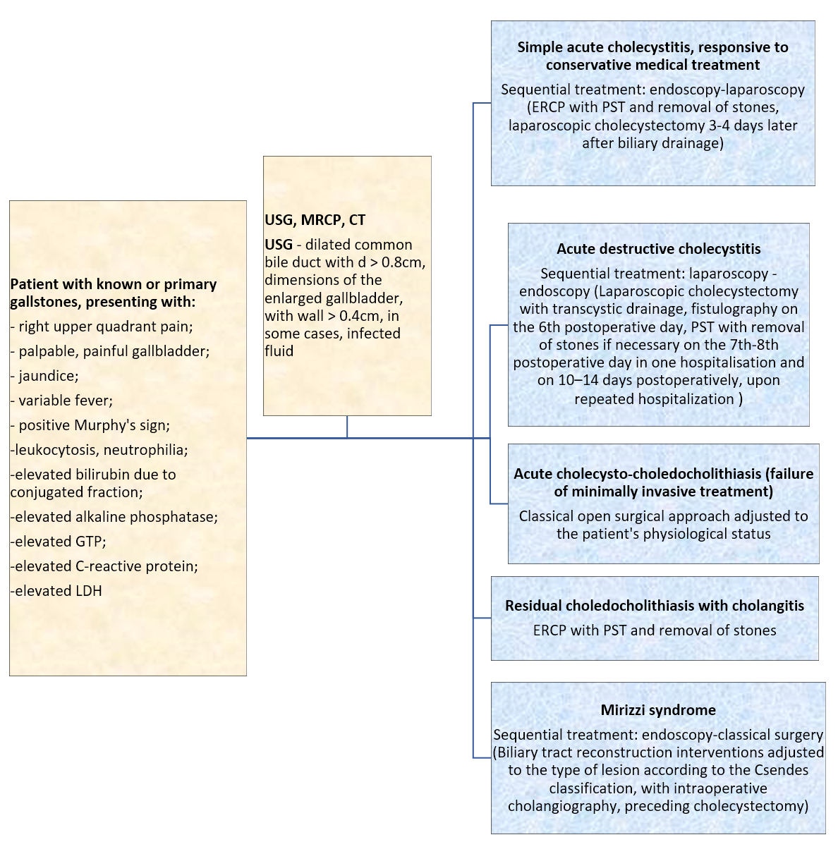
Using the existing recommendations for the approach to patients with inflammatory complications and cholestatic jaundice, alongside our own clinical results, we opted for a sequential treatment strategy. The staging of sequential surgical treatment for obstructive jaundice and infectious inflammatory lesions was differentiated in the study group based on two important factors: the severity of the infectious process and the stable or recurrent nature of the cholestatic jaundice. In developing the algorithm, our primary objective was the improvement of bile flow, with a strong preference for internal biliary drainage methods; however, in cases of a severe infection, external biliary drainage at the first stage was preferential. The diagnostic and treatment strategy algorithm is shown in Figure 3.
Minimally invasive surgical approaches (1. endoscopy-laparoscopy and 2. laparoscopy-endoscopy) were utilized, while classical open surgery was performed in cases where minimally invasive maneuvers failed. Open surgery remained frequently required in this patient cohort. Clinical and diagnostic evaluations of the patient's general condition and morbidity risk were the fundamental pillars for differentiating the applied treatment tactics and techniques.
Internal decompression at the first stage via endoscopic papillosphincterotomy (PST), with or without stone removal within the first 48–72 hours of hospitalization, was used in 16 (23.9%) cases of simple acute cholecystitis responsive to conservative medical treatment. This was followed by laparoscopic cholecystectomy in the second stage, within 3–4 days after biliary drainage. The average length of hospital stay for these patients was 10.18 ± 1.2 days.
In 31 (46.3%) cases of destructive acute cholecystitis and severe II grade cholangitis, first-stage decompression was achieved via cholecystectomy with external drainage of the biliary tree. The presence of acute destructive cholecystitis, confirmed by laboratory and imaging findings, necessitated an urgent surgical intervention to remove the septic focus and provide external drainage of the infected biliary tract. Laparoscopic cholecystectomy with Halsted (transcystic) drainage was successful in 10 (32.3%) cases, whereas open surgery – cholecystectomy, choledochotomy with stone extraction and Kehr (T-tube) drainage – was applied in 21 (67.7%) patients.
Fistulo-cholangiography, performed on the 5th to 7th postoperative day, revealed biliary tract disorders that required PST in both surgical groups. Specifically, PST was necessary for 17 (80.9%) patients operated on via the classical open approach, and for 8 (80%) patients who underwent laparoscopic treatment. Among the laparoscopic group, 6 (60%) patients also presented with choledocholithiasis. To address this, these patients underwent a second-stage ERCP with PST and successful stone extraction 14–20 days after their initial laparoscopic surgery, during a subsequent hospital readmission.
Compromised biliary passage was identified in 25 (80.6%) of the 31 operated patients. In this cohort, postoperative fistulography was useful for identifying patients requiring further biliary intervention. External biliary drainage provided temporary decompression; restoration of internal biliary flow, when indicated, was subsequently achieved using contrast assessment and endoscopic intervention. The mean length of hospital stay was 14.22 ± 1.3 days for the laparoscopy-endoscopy sequence (including readmissions) and 18.56 ± 2.3 days for the laparotomy-endoscopy sequence.
In 7 (10.4%) cases of residual choledocholithiasis and severe cholangitis, complete clearance of stones and resolution of the jaundice syndrome were achieved by PST with stone extraction. This was combined with endoscopic nasobiliary drainage in 3 cases, and with the placement of an internal biliary stent in 1 case. The average length of hospital stay was 10.1 ± 0.8 days.
All patients with confirmed Mirizzi syndrome presented with a long-standing history of gallstones, in our study, where the average history of gallstones was estimated at 10.1 ± 1.8 years, associated with characteristic biliary colic, and evidence of cholestatic jaundice associated with recurrent cholangitis. The preferred preoperative diagnostic modality was magnetic resonance cholangiopancreatography (MRCP). These patients were managed sequentially with an endoscopy-laparotomy approach. During the first stage, ERCP served as a diagnostic tool and facilitated biliary drainage via PST, which was combined with Bailey's nasobiliary drainage in 3 (23%) cases complicated by severe cholangitis. In the second stage, patients underwent biliary tract reconstruction adjusted to the type of obstruction, which was assessed using intraoperative cholangiography in 10 (76.9%) cases. The specific surgical procedure was selected depending on the type of lesion, according to the Csendes classification (1989): type I was found in 2 (15.4%) cases, type II – in 4 (30.8%), type III – in 5 (38.5%), type IV – in 2 (15.4%) cases. For the 2 (15.4%) type I cases stone removal was performed via cholecystectomy with Kehr drainage. For the 4 (30.8%) type II cases, stone removal with fistular defect closure was performed, preferably utilizing a sparing gallbladder wall flap and Kehr biliary drainage. In types III and IV, biliary-digestive anastomoses were applied: cholecystectomy, choledocholithotomy with stone extraction and choledochoduodenal anastomosis in 4 (30.8%) cases; and cholecystectomy, choledocholithotomy with stone extraction, and Roux-en-Y choledochojejunal anastomosis – in 3 (23.1%) cases. The average length of hospital stay was 21.32 ± 9 days.
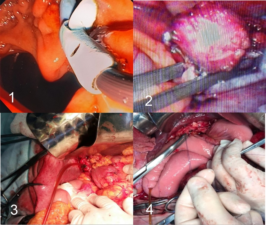
Intra- and postoperative complications observed in the evaluated group included: allergic reaction to contrast media during ERCP (n = 1); acute pancreatitis following ERCP with PST (n = 1); postoperative pneumonia (n = 1); postoperative wound suppuration (n = 4); dehiscence of the biliary-digestive anastomosis (n = 1), subhepatic biloma, no extension, which required relaparotomy with the creation of a new anastomosis and external transduodenal drainage of the biliary tract. Eight complications were reported in the cohort of 67 patients; no deaths occurred. Treatment options in infectious complications are shown in Figure 4.
Incidence of obstructive jaundice syndrome in diseases of the hepato-pancreatobiliary system of 2.6%–23.7% included cholestatic jaundice in complications of gallstone disease, with the incidence of 10%–15% of cases [1, 2, 3], but the progressive complications produced by the associated cholestasis pose a significant risk in the patient's prognosis. Diagnostic errors during the investigation stage contribute to the early development of severe complications in 12%–38% of cases — such as purulent cholangitis, liver abscesses, biliary sepsis and hemorrhage — which lead to a progressive hepatocellular insufficiency with a mortality rate of 14%–27% [12].
Treatment strategies reported in the literature emphasize timely intervention in patients with biliary infection and obstruction. The specific approach varies according to clinical severity, anatomy, local expertise, and available resources, with an increasing role for laparoscopic and endoscopic techniques [2, 5, 7]. The range of available tactics and techniques reflects the need to individualize management rather than a single approach applicable to all patients [5, 6, 7, 13].
Biliary decompression is an important component of management when obstruction is accompanied by cholangitis or persistent cholestasis. Depending on disease severity and local expertise, definitive treatment may be performed during the same admission or as part of a staged approach [4, 5, 9, 10, 11]. The choice between single-stage and sequential strategies should consider the patient’s clinical condition, comorbidities, anatomy, and the availability of endoscopic and laparoscopic expertise.
Sequential treatment with initial biliary decompression is one of the available strategies for selected patients with obstructive jaundice and biliary infection. The optimal timing of definitive gallstone treatment varies according to clinical severity and the intervention performed. Contemporary recommendations support early source control and biliary drainage when indicated, while the timing of cholecystectomy should be individualized according to the patient’s condition and operative risk [10, 11].
Current literature demonstrates a role for both classical open and minimally invasive surgery, highlighting the need to individualize surgical tactics on a case-by-case basis [6, 9, 14, 15, 16]. An important consideration in these patients is that the severity of the local inflammatory process near the common bile duct (CBD) may render endoscopic PST via ERCP impossible; in such scenarios, intraoperative cholangiography during classical or laparoscopic surgery serves as a necessary alternative [14].
The timing of surgery should be determined by the severity of inflammation, the presence of cholangitis or biliary obstruction, the patient’s physiological status, and available expertise. Current guidelines favor early laparoscopic cholecystectomy for suitable patients with acute cholecystitis, while drainage followed by delayed surgery remains an option when early surgery is not appropriate [11]. In the historical cohort analysed in this study, delayed or staged procedures were used selectively according to clinical status and local treatment possibilities.
In the present cohort, treatment was staged according to the morphological form of inflammation and the patient’s clinical condition. In simple forms responsive to conservative treatment, an endoscopy-laparoscopy sequence was used, with endoscopic PST (with or without stone extraction) followed by cholecystectomy. In destructive cholecystitis and severe cholangitis, external biliary drainage associated with laparoscopic or open surgery was used first, with subsequent endoscopic intervention when indicated by fistulocholangiography. Eight complications were reported, with no deaths; however, the descriptive design and heterogeneity of the subgroups do not allow conclusions regarding superiority over alternative strategies.
Mirizzi syndrome is reported to have an incidence of 0.7% to 1.4% among patients undergoing cholecystectomy [18, 19], and is characteristic of elderly patients with a prolonged history of gallstone disease. The typical clinical presentation included repeated bouts of biliary colic associated with cholestasis and recurrent cholangitis — symptoms observed in all examined patients. Preoperatively, the diagnosis is routinely suspected using USG, MRCP and ERCP, a finding supported in this study by assessing the diagnostic efficacy of MRCP and ERCP. However, to accurately evaluate the lesion type according to the Csendes classification, intraoperative cholangiography prior to gallbladder dissection was utilized and is highly recommended to determine the topographic features of the fistula defect [18, 19, 20].
Patient management methods described in the literature include both laparoscopic and open surgery. While laparoscopic surgery is preferable for Mirizzi syndrome types I and II, it carries a higher risk of biliary tract injury. Studies report baseline injury rates of 0.3% to 1.0% during these procedures; however, in emergency interventions lacking complete preoperative diagnostic imaging, this risk can increase to 17% [17, 18, 19, 20].
In the staged treatment approach, we opted for first-stage decompression through PST and biliary drainage as needed. In the second stage, we performed biliary tract reconstruction tailored to the specific type, morphological features, and topography of the fistula. Among the severe postoperative complications observed one particular case merits attention: a 78-year-old patient with concomitant grade III heart failure and type III Mirizzi syndrome. On the third postoperative day, the patient developed dehiscence of the choledochoduodenal anastomosis, leading to localized biliary peritonitis, which was evidenced by bile exteriorizing through the surgical drains. The patient underwent emergency relaparotomy to restore the choledochoduodenal anastomosis by suturing the anastomotic defect alongside nasobiliary drainage. The biliary drain was successfully removed 12 days after relaparotomy.
1. The use of minimally invasive techniques within a sequential treatment strategy was associated with eight reported complications and no deaths in the studied cohort; the length of hospital stay varied according to the treatment sequence and clinical subgroup.
2. In the studied cohort, the timing and type of intervention were individualized according to the morphological form of the disease and the patient's clinical condition, with biliary decompression prioritized in the first stage of sequential management.
Omarov N, Aimagambetov M, Auyenov M, Abdrakhmanov S, Bulegenov T, Noso Y. Obstructive jaundice syndrome of benign genesis and its surgical treatment. Int J Health Sci. 2022;6(suppl 7):3133-3151. doi:10.53730/ijhs.v6nS7.12118
Taylor T, Wheatley M, Gupta N, Nusbaum J. Jaundice in the emergency department: meeting the challenges of diagnosis and treatment [digest]. Emerg Med Pract. 2018;20(suppl 4):1-2.
Shabanzadeh DM. Incidence of gallstone disease and complications. Curr Opin Gastroenterol. 2018;34(2):81-89. doi:10.1097/MOG.0000000000000418
Attasaranya S, Fogel EL, Lehman GA. Choledocholithiasis, ascending cholangitis, and gallstone pancreatitis. Med Clin North Am. 2008;92(4):925-960, x. doi:10.1016/j.mcna.2008.03.001
Barkun JS, Chaudhury P, Barkun AN. Jaundice. In: ACS Surgery: Principles and Practice. 2006. https://www.slideshare.net/slideshow/acs0503-jaundice-2006/4839276
Modha K. Clinical approach to patients with obstructive jaundice. Tech Vasc Interv Radiol. 2015;18(4):197-200. doi:10.1053/j.tvir.2015.07.002
Cuschieri A, Lezoche E, Morino M, et al. E.A.E.S. multicenter prospective randomized trial comparing two-stage vs single-stage management of patients with gallstone disease and ductal calculi. Surg Endosc. 1999;13(10):952-957. doi:10.1007/s004649901145
Wang Q, Gurusamy KS, Lin H, Xie X, Wang C. Preoperative biliary drainage for obstructive jaundice. Cochrane Database Syst Rev. 2008;(3):CD005444. doi:10.1002/14651858.CD005444.pub2
Shi H, Wan J, Xu SP, Liao L. A retrospective analysis of clinical characteristics and mortality risks in elderly patients with acute cholecystitis and cholangitis. Zhonghua Nei Ke Za Zhi. 2019;58(6):415-418. doi:10.3760/cma.j.issn.0578-1426.2019.06.003 [In Chinese].
Clayton ESJ, Connor S, Alexakis N, Leandros E. Meta-analysis of endoscopy and surgery versus surgery alone for common bile duct stones with the gallbladder in situ. Br J Surg. 2006;93(10):1185-1191. doi:10.1002/bjs.5568
Miura F, Okamoto K, Takada T, et al. Tokyo Guidelines 2018: initial management of acute biliary infection and flowchart for acute cholangitis. J Hepatobiliary Pancreat Sci. 2018;25(1):31-40. doi:10.1002/jhbp.509
Okamoto K, Suzuki K, Takada T, et al. Tokyo Guidelines 2018: flowchart for the management of acute cholecystitis. J Hepatobiliary Pancreat Sci. 2018;25(1):55-72. doi:10.1002/jhbp.516
Kodirov MS, Turakulov UN, Kodirov SN. Factors leading of obstructive jaundice syndrome and its complications in elderly patient. Am J Med Med Sci. 2024;14(11):2711-2715. doi:10.5923/j.ajmms.20241411.06
Strelțov L. Colestatic cholemia in inflammatory complications of biliary lithiasis, stage treatment options. Revista de Științe ale Sănătății din Moldova. 2022;3(29):316. [Conference abstract].
Salminen P, Laine S, Gullichsen R. Severe and fatal complications after ERCP: analysis of 2555 procedures in a single experienced center. Surg Endosc. 2008;22(9):1965-1970. doi:10.1007/s00464-007-9711-0
Atanasova N. The role of open surgery in the treatment of benign mechanical jaundice: a retrospective single-center study. Scripta Scientifica Medica. 2025;57(suppl 2):30-35. doi:10.14748/kzcnz423
Liu JJ, Sun YM, Xu Y, Mei HW, Guo W, Li ZL. Pathophysiological consequences and treatment strategy of obstructive jaundice. World J Gastrointest Surg. 2023;15(7):1262-1276. doi:10.4240/wjgs.v15.i7.1262
Bellamlih H, Bouimetarhan L, En-Nouali H, et al. Le syndrome de Mirizzi: une cause rare de l’obstruction des voies biliaires: à propos d’un cas et revue de littérature. Pan Afr Med J. 2017;27:45. doi:10.11604/pamj.2017.27.45.12469 (In French).
Hotineanu A, Ferdohleb A, Cucu I. Sindromul Mirizzi: particularități de diagnostic și tratament chirurgical. Chirurgia. 2022;117(suppl 1):S126-S127. (In Romanian).
Streltov L. Sindromul Mirizzi: considerații diagnostice și terapeutice. Bul Acad Științe Mold Științe Med. 2016;52(3):82-87. https://bulmed.md/bulmed/article/view/2639 (In Romanian).